Overview
Achilles tendon ruptures may be divided into full thickness ("total") and partial thickness ruptures. Total ruptures usually occur in formerly active athletes (average age 40) who resume sport activity after having been away from it for some time. In these cases, degenerative changes have weakened the tendon so much that sudden, forceful loading of the tendon causes it to tear. To some extent, these changes in the tendon could have been prevented by regular physical activity. In most cases, the injury mechanism is a strong activation of the posterior lower leg musculature, eccentrically overloading the tendon. A typical mechanism of injury involves pushing off hard with the weight-bearing foot while the knee is extended (e.g., running uphill) or sudden, unexpected dorsal extension of the ankle with reflex contraction of the calf musculature (e.g., falling down into a hole).
Causes
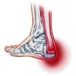
The Achilles tendon is a strong bands of fibrous connective tissue that attaches the calf muscle to the heel bone. When the muscle contracts, the tendon transmits the power of this contraction to the heel bone, producing movement. The Achilles tendon ruptures because the load applied to it is greater than the tendon's ability to withstand that load. This usually occurs as a result of a sudden, quick movement where there is a forceful stretch of the tendon or a contraction of the muscles eg: jumping, sprinting, or pushing off to serve in tennis. This occurs most often in sports that require a lot of stopping and starting (acceleration-deceleration sports) such as tennis, basketball, netball and squash. The Achilles tendon is on average 15cm in length. Most ruptures occur 2-6cm above where the tendon inserts into the heel bone. This is the narrowest portion of the Achilles tendon and is also the area with the poorest blood supply. achilles tendon rupture is most common when the muscles and tendon have not been adequately stretched and warmed up prior to exercise, or when the muscles are fatigued. the Achilles tendon has a poor blood supply, which makes it susceptible to injury and slow to heal after injury. During exercise the amount of blood able to travel to the tendon is decreased, further increasing the risk of rupture. Most experts agree that there are no warning signs of an impending rupture. However, frequent episodes of Achilles tendonitis (tendon inflammation) can weaken the tendon and make it more susceptible to rupture.
Symptoms
Tendon strain or tendon inflammation (tendonitis) can occur from tendon injury or overuse and can lead to a rupture. Call your doctor if you have signs of minor tendon problems. Minor tenderness and possible swelling increases with activity. There is usually no specific event causing sudden pain and no obvious gap in the tendon. You can still walk or stand on your toes. Acute calf pain and swelling can indicate a tear or partial tear of the Achilles tendon where it meets the calf muscle. You may still be able to use that foot to walk, but you will need to see a specialist such as an orthopedic surgeon. Surgery is not usually done for partial tears. Sometimes special heel pads or orthotics in your shoes may help. Follow up with your doctor to check for tendonitis or strain before resuming activity, because both can increase the risk of tendon rupture. Any acute injury causing pain, swelling, and difficulty with weight-bearing activities such as standing and walking may indicate you have a tear in your Achilles tendon. Seek prompt medical attention from your doctor or emergency department. Do not delay! Early treatment results in better outcome. If you have any question or uncertainty, get it checked.
Diagnosis
The diagnosis of an Achilles tendon rupture can be made easily by an orthopedic surgeon. The defect in the tendon is easy to see and to palpate. No x-ray, MRI or other tests are necessary.
Non Surgical Treatment
The other option is to allow your tendon to heal without surgery. In this case, you also need to wear a cast, splint, walking boot, or brace for 6-8 weeks. You also may have different exercises to do. If you are less active or have a chronic illness that prevents surgery, this option may be better for you.
Surgical Treatment
Most published reports on surgical treatment fall into 3 different surgical approach categories that include the following: direct open, minimally invasive, and percutaneous. In multiple studies surgical treatment has demonstrated a lower rate of re-rupture compared to nonoperative treatment, but surgical treatment is associated with a higher rate of wound healing problems, infection, postoperative pain, adhesions, and nerve damage. Most commonly the direct open approach involves a 10- to 18-cm posteromedial incision. The minimally invasive approach has a 3- to 10-cm incision, and the percutaneous approach involves repairing the tendon through multiple small incisions. As with nonsurgical treatment there exists wide variation in the reported literature regarding postoperative treatment protocols. Multiple comparative studies have been published comparing different surgical approaches, repair methods, or postoperative treatment protocols.
Achilles tendon ruptures may be divided into full thickness ("total") and partial thickness ruptures. Total ruptures usually occur in formerly active athletes (average age 40) who resume sport activity after having been away from it for some time. In these cases, degenerative changes have weakened the tendon so much that sudden, forceful loading of the tendon causes it to tear. To some extent, these changes in the tendon could have been prevented by regular physical activity. In most cases, the injury mechanism is a strong activation of the posterior lower leg musculature, eccentrically overloading the tendon. A typical mechanism of injury involves pushing off hard with the weight-bearing foot while the knee is extended (e.g., running uphill) or sudden, unexpected dorsal extension of the ankle with reflex contraction of the calf musculature (e.g., falling down into a hole).
Causes
The Achilles tendon is a strong bands of fibrous connective tissue that attaches the calf muscle to the heel bone. When the muscle contracts, the tendon transmits the power of this contraction to the heel bone, producing movement. The Achilles tendon ruptures because the load applied to it is greater than the tendon's ability to withstand that load. This usually occurs as a result of a sudden, quick movement where there is a forceful stretch of the tendon or a contraction of the muscles eg: jumping, sprinting, or pushing off to serve in tennis. This occurs most often in sports that require a lot of stopping and starting (acceleration-deceleration sports) such as tennis, basketball, netball and squash. The Achilles tendon is on average 15cm in length. Most ruptures occur 2-6cm above where the tendon inserts into the heel bone. This is the narrowest portion of the Achilles tendon and is also the area with the poorest blood supply. achilles tendon rupture is most common when the muscles and tendon have not been adequately stretched and warmed up prior to exercise, or when the muscles are fatigued. the Achilles tendon has a poor blood supply, which makes it susceptible to injury and slow to heal after injury. During exercise the amount of blood able to travel to the tendon is decreased, further increasing the risk of rupture. Most experts agree that there are no warning signs of an impending rupture. However, frequent episodes of Achilles tendonitis (tendon inflammation) can weaken the tendon and make it more susceptible to rupture.
Symptoms
Tendon strain or tendon inflammation (tendonitis) can occur from tendon injury or overuse and can lead to a rupture. Call your doctor if you have signs of minor tendon problems. Minor tenderness and possible swelling increases with activity. There is usually no specific event causing sudden pain and no obvious gap in the tendon. You can still walk or stand on your toes. Acute calf pain and swelling can indicate a tear or partial tear of the Achilles tendon where it meets the calf muscle. You may still be able to use that foot to walk, but you will need to see a specialist such as an orthopedic surgeon. Surgery is not usually done for partial tears. Sometimes special heel pads or orthotics in your shoes may help. Follow up with your doctor to check for tendonitis or strain before resuming activity, because both can increase the risk of tendon rupture. Any acute injury causing pain, swelling, and difficulty with weight-bearing activities such as standing and walking may indicate you have a tear in your Achilles tendon. Seek prompt medical attention from your doctor or emergency department. Do not delay! Early treatment results in better outcome. If you have any question or uncertainty, get it checked.
Diagnosis
The diagnosis of an Achilles tendon rupture can be made easily by an orthopedic surgeon. The defect in the tendon is easy to see and to palpate. No x-ray, MRI or other tests are necessary.
Non Surgical Treatment
The other option is to allow your tendon to heal without surgery. In this case, you also need to wear a cast, splint, walking boot, or brace for 6-8 weeks. You also may have different exercises to do. If you are less active or have a chronic illness that prevents surgery, this option may be better for you.
Surgical Treatment
Most published reports on surgical treatment fall into 3 different surgical approach categories that include the following: direct open, minimally invasive, and percutaneous. In multiple studies surgical treatment has demonstrated a lower rate of re-rupture compared to nonoperative treatment, but surgical treatment is associated with a higher rate of wound healing problems, infection, postoperative pain, adhesions, and nerve damage. Most commonly the direct open approach involves a 10- to 18-cm posteromedial incision. The minimally invasive approach has a 3- to 10-cm incision, and the percutaneous approach involves repairing the tendon through multiple small incisions. As with nonsurgical treatment there exists wide variation in the reported literature regarding postoperative treatment protocols. Multiple comparative studies have been published comparing different surgical approaches, repair methods, or postoperative treatment protocols.
 RSS Feed
